Pesquisadores aceleram técnicas de imagem para capturar estruturas de moléculas pequenas
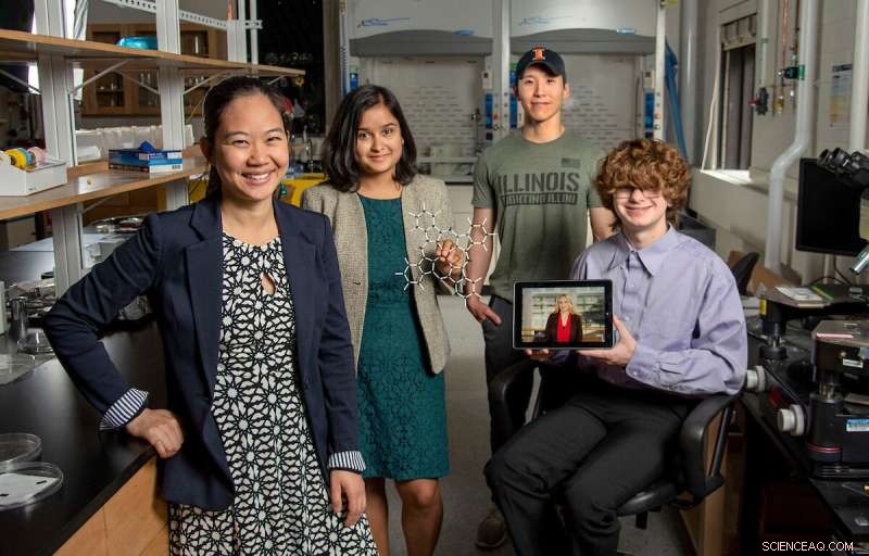
Pinshane Huang, à esquerda, junta-se a Priti Kharel, segundo da esquerda, Justin Bae e Patrick Carmichael, à direita, no Laboratório de Pesquisa de Materiais. Na foto no tablet está Blanka Janicek. Crédito:Heather Coit/The Grainger College of Engineering
Um esforço de pesquisa da Universidade de Illinois Urbana-Champaign, liderado por Pinshane Huang, está acelerando as técnicas de imagem para visualizar claramente estruturas de pequenas moléculas – um processo antes considerado impossível. Sua descoberta libera um potencial infinito para melhorar as aplicações da vida cotidiana – de plásticos a produtos farmacêuticos.
O professor associado do Departamento de Ciência e Engenharia de Materiais juntou-se aos autores co-líderes Blanka Janicek, ex-aluno de 21 e pós-doutorado no Laboratório Nacional Lawrence Berkeley em Berkeley, Califórnia, e Priti Kharel, estudante de pós-graduação do Departamento de Química, para provar a metodologia que permite aos pesquisadores visualizar pequenas estruturas moleculares e acelerar as técnicas de imagem atuais.
Coautores adicionais incluem o estudante de pós-graduação Sang hyun Bae e os graduandos Patrick Carmichael e Amanda Loutris. Sua pesquisa revisada por pares foi publicada recentemente em
Nano Letters .
Os esforços da equipe expõem a estrutura atômica da molécula, permitindo que os pesquisadores entendam como ela reage, aprendam seus processos químicos e vejam como sintetizar seus compostos químicos.
"A estrutura de uma molécula é tão fundamental para sua função", disse Huang. "O que fizemos em nosso trabalho é tornar possível ver essa estrutura diretamente."
A capacidade de ver a estrutura de uma pequena molécula é vital. Kharel compartilha o quão vital é dando o exemplo de um medicamento conhecido como talidomida.
Descoberta nos anos 60, a talidomida foi prescrita para mulheres grávidas para tratar enjôos matinais e, mais tarde, descobriu-se que causava graves defeitos congênitos ou, em alguns casos, até a morte.
O que deu errado? A droga tinha estruturas moleculares mistas, uma responsável pelo tratamento do enjoo matinal e a outra, infelizmente, causando efeitos adversos devastadores ao feto.
A necessidade de uma ciência proativa, não reativa, incitou Huang e seus alunos a buscarem esse esforço de pesquisa que originalmente começou com pura curiosidade.
"É tão crucial determinar com precisão as estruturas dessas moléculas", disse Kharel.
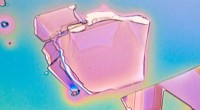
Normalmente, as estruturas moleculares são determinadas com técnicas indiretas, uma abordagem demorada e difícil que usa ressonância magnética nuclear ou difração de raios X. Pior ainda, métodos indiretos podem produzir estruturas incorretas que dão aos cientistas uma compreensão errada da composição de uma molécula por décadas. A ambiguidade em torno das estruturas das pequenas moléculas pode ser eliminada usando métodos de imagem direta.
In the last decade, Huang has seen significant advancements in cryogenic electron microscopy technology, where biologists freeze the large molecules to capture high-quality images of their structures.
"The question that I had was:What's keeping them from doing that same thing for small molecules?" Huang said. "If we could do that, you might be able to solve the structure (and) figure out how to synthesize a natural compound that a plant or animal makes. This could turn out to be really important, like a great disease-fighter," Huang said.
The challenge is that small molecules are often 100 or even 1,000 times smaller than large molecules, making their structures difficult to detect.
Determined, Huang's students began using existing large molecule methodology as a starting point for developing imaging techniques to make the small molecules' structures appear.
Unlike large molecules, the imaging signals from small molecules are easily overwhelmed by their surroundings. Instead of using ice, which typically serves as a layer of protection from the harsh environment of the electron microscope, the team devised another plan for keeping the small molecules' structures intact.
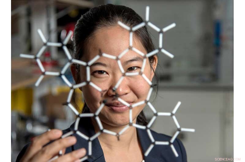
Pinshane Huang and her researchers discovered how to view structures in molecules, which opens a whole new realm of scientific possibilities. Credit:Heather Coit/The Grainger College of Engineering
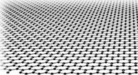
How can you temper a molecule's environment? By using graphene.
Graphene, a single layer of carbon atoms that form a tight, hexagon-shaped honeycomb lattice, dissipates damaging reactions during imaging.
Stabilizing the small molecule's environment was only one issue the Illinois researchers had to manage. The team also had to limit its use of electrons, as low as one-millionth the number of electrons normally used, to illuminate the molecules.
Low doses of electrons ensure that the molecules are still moving enough for the researchers to capture an image.
"The way I like to think about it is the molecule doesn't like to be bombarded by higher-energy electrons, but we need to do that to be able to see the structure, and graphene helps dissipate some of that charge away from the molecule so that we can actually get a nice image of it," Janicek said.
Unfortunately, once captured, the molecules were nearly invisible in the image.
"When they take a low-dose image, it initially looks like noise or TV static—almost like nothing is there," Huang said.
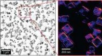
The trick was to isolate the atomic structures from that noise by using a Fourier transform—a mathematical function that breaks down the small molecule's image—to see its spatial frequency.
"We took images of hundreds of thousands of molecules and added them together to build a single, clear image," Kharel said.
This averaging approach allowed the team to create crisp images of the molecules' atoms without damaging the integrity of any individual molecule.
"Month after month, week after week, our resolution improved," Huang said. "And then one day, my students came in and showed me the individual carbon atoms—that's a major achievement. And of course, it comes after all this deep knowledge that they have gained to design an imaging experiment and how to unlock data from what looks like nothing."
This collective discovery is paving the way for many more structural molecule imaging findings.
"There's been this whole field of small molecules that have been left out in the cold, so to speak. We're shining a light on how do we get there as a field? How do we make this thing that for us right now is so hard?" Huang said. "One day it won't be—that's the hope."
The Illinois researchers' efforts are the first big step in turning that dream into reality.
"One day, this will be the way we solve the structure of a small molecule," Huang said. "People will simply throw the molecule in the electron microscope, take a picture and be done."
That dream inspires Huang and her Illinois team to keep the course.
"That's potentially life-changing, and we've made it exist," Huang said. "We haven't yet made it easy, but imaging techniques like this will change so much of science and technology."
+ Explorar mais ESR-STM em moléculas únicas e estruturas baseadas em moléculas