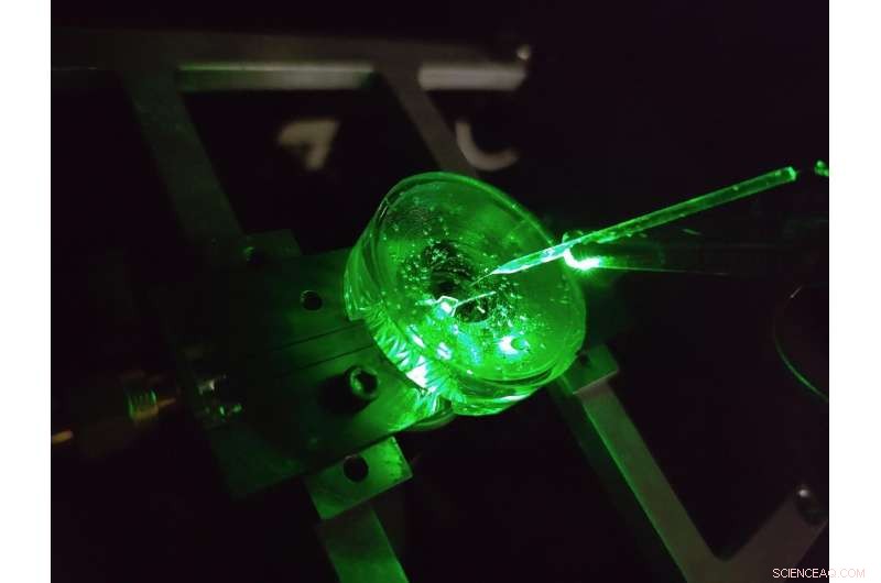
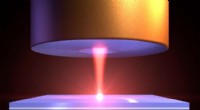
Um protótipo de um microscópio de imagem de voltagem de diamante construído por físicos da Universidade de Melbourne. Um pequeno eletrodo é suspenso acima do chip de diamante para testar o desempenho do dispositivo. Um laser verde brilhou de baixo para fornecer excitação de fluorescência ao chip. Crédito:Autor fornecido, Universidade de Melbourne
O cérebro é sem dúvida uma das estruturas mais complexas do universo conhecido. Avanços contínuos em nossa compreensão do cérebro e nossa capacidade de tratar efetivamente uma série de doenças neurológicas dependem da sondagem dos microcircuitos neurais do cérebro com detalhes cada vez maiores.
Uma classe de métodos para estudar circuitos neurais é chamada de imagem de tensão. Essas técnicas nos permitem ver a voltagem gerada pelos neurônios de disparo do nosso cérebro – nos dizendo como as redes de neurônios se desenvolvem, funcionam e mudam ao longo do tempo.
Hoje, a geração de imagens de voltagem de neurônios cultivados é realizada usando densos arranjos de eletrodos nos quais as células são cultivadas (ou cultivadas), ou aplicando corantes emissores de luz que respondem opticamente a mudanças na voltagem na superfície da célula.
Mas o nível de detalhe que podemos ver usando essas técnicas é restrito.
Os menores eletrodos não conseguem distinguir neurônios individuais de forma confiável, com cerca de 20 milionésimos de metro de diâmetro, para não falar da densa rede de conexões em nanoescala que se forma entre eles, e nenhum avanço tecnológico significativo foi feito nessa área por mais de duas décadas.
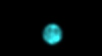
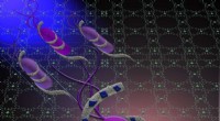
Além disso, cada eletrodo requer sua própria conexão com fio e amplificador, colocando limitações significativas no número de eletrodos que podem ser medidos simultaneamente. Um eletrodo minúsculo, com oito milionésimos de metro de diâmetro, é usado para injetar localmente uma nuvem de carga elétrica em um líquido colocado no topo do lasca de diamante. A fluorescência do diamante espelha a difusão desta carga através do líquido em tempo real. Crédito:Autor fornecido Os corantes podem superar essas limitações ao visualizar a voltagem sem fio como luz – isso significa que a eletrônica complexa pode ser situada longe das células dentro de uma câmera.
O resultado é alta resolução em grandes áreas, capaz de distinguir cada neurônio individual em uma grande rede. Mas há limitações aqui também, as respostas de voltagem dos corantes de última geração são lentas e instáveis.
Nossa pesquisa recente publicada na Nature Photonics , explora um novo tipo de plataforma de imagem de alta velocidade, alta resolução e tensão escalável criada com o objetivo de superar essas limitações - um microscópio de imagem de voltagem de diamante.
Desenvolvido por uma equipe de físicos da Universidade de Melbourne e da Universidade RMIT, o dispositivo usa um sensor baseado em diamante que converte sinais de tensão em sua superfície diretamente em sinais ópticos – isso significa que podemos ver a atividade elétrica enquanto ela acontece.
A conversão usa as propriedades de um defeito em escala atômica na estrutura cristalina do diamante conhecido como vacância de nitrogênio (NV).
NV defects can be engineered by bombarding the diamond with a nitrogen ion beam using a special type of particle accelerator. The fabrication of the sensor begins with using this process to create a high-density, ultra-thin layer of NV defects close to the diamond's surface.
You can think of each NV defect as a bucket that holds up to two electrons. When this bucket is empty, the NV defect is dark. With one electron, the NV defect emits orange light when illuminated by a laser—this property is known as fluorescence. With two electrons, the color of the fluorescence becomes red. As the voltage in a conductive solution is uniformly varied, the brightness of light emitted by the diamond chip follows with a near instantaneous response. Here, the diamond surface has been patterned into an array of nanopillars to increase the detected light signal. Credit:Author provided A previously discovered property of NV defects is that the number of electrons they hold—and the resulting fluorescence—can be controlled with a voltage. Unlike dyes, the voltage response of an NV defect is very fast and stable.
Our research aims to overcome the challenge of making this effect sensitive enough to image neuronal activity.
On the diamond's surface, the crystal structure ends with a layer one atom thick, made up of hydrogen and oxygen atoms. The NV defects closest to the surface are the most sensitive to changes in voltage outside the diamond, but they are also highly sensitive to the atomic makeup of the surface layer.
Too much hydrogen and the NVs are so dark that the optical signals we are looking for cannot be seen. Too little hydrogen and the NVs are so bright that the small signals we are after are completely washed out.
So, there's a "Goldilocks' zone" for voltage imaging, where the surface has just the right amount of hydrogen.
To reach this zone, our team developed an electrochemical method for removing hydrogen in a controlled way. By doing this, we've managed to achieve voltage sensitivities two orders of magnitude better than what has been previously reported.
We tested our sensor in salty water using a microscopic wire 10-times thinner than a human hair. By applying a current, the wire can produce a small cloud of charge in the water above the diamond. The formation and subsequent diffusion of this charge cloud produces small voltages at the diamond surface.
By capturing these voltages through a high-speed recording of the NV fluorescence, we can determine the speed, sensitivity and resolution of our diamond imaging chip.
We were able to further boost sensitivity by patterning the diamond's surface into 'nanopillars'—conical structures with the NV centers embedded in their tips. These pillars funnel the light emitted by the NVs towards the camera, dramatically increasing the amount of signal we can collect.
With the development of the diamond voltage imaging microscope for detecting neuronal activity, the next step is the recording of activity from cultured neurons in vitro—these are experiments on cells grown outside their normal biological context, otherwise known as test-tube or petri-dish experiments.
What differentiates this technology from existing state-of-the-art in vitro techniques is the combination of high spatial resolution (on the order of a millionth of a meter or less), large spatial scale (a few millimeters in each direction—which for a network of neurons in mammals is quite vast), and complete stability over time.
No other existing system can simultaneously offer these three qualities, and it's this combination that will allow our made-in-Melbourne technology to make a valuable contribution to the work of neuroscientists and neuropharmacologists globally.
Our system will aid these researchers in pursuing both fundamental knowledge and the next generation of treatments for neurological and neurodegenerative diseases. + Explorar mais
New method enables long-lasting imaging of rapid brain activity in individual cells deep in the cortex